Diagnosing a fungal infection of the outermost layer of the eye is a major bottleneck for the treatment of this widespread and impairing condition. Currently, diagnostics are time-consuming and not available for everybody. A test that can be used outside of medical centres, close to patients and giving results within hours would help many people in underserved regions. Once identified early and correctly, it only takes the right eye drops and time for the infection to heal.

Bringing diagnostics of fungal keratitis closer to patients
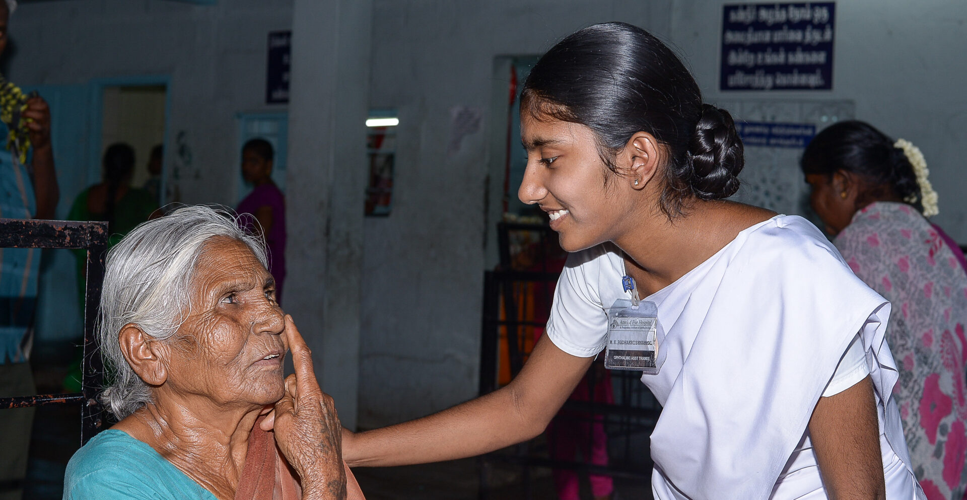
A slight injury of the outermost layer of the eye, the cornea, is enough to increase the risk of corneal infection by fungi. The condition is called fungal keratitis and occurs worldwide, but it is much more frequent in tropics and subtropics. Such fungal infection of the cornea initially leads to blurred vision, painful eyes and increased tears or discharge. Without treatment though, the resulting visual impairment can be severe, making it a major cause of blindness especially in low- and middle-income countries.
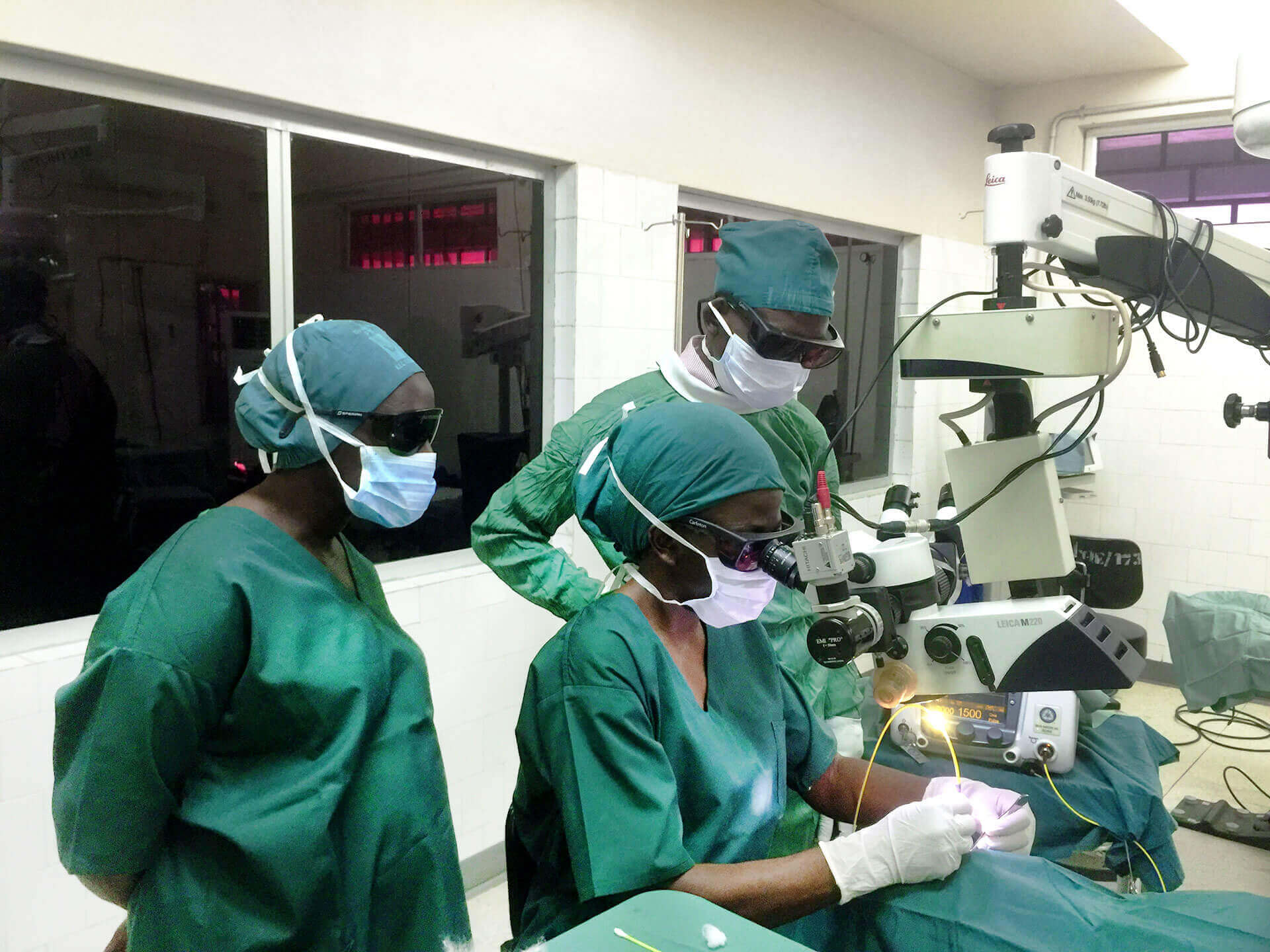
In many places a major bottleneck in managing fungal keratitis is the diagnosis. Often primary care physicians or general ophthalmologists refer people to cornea specialists. The current diagnostic tests take time, need expensive equipment as well as skilled technicians and often lack sensitivity. In low- and middle-income countries, patients at risk of infection are often agricultural workers who experience trauma to the cornea by handling plant materials. They frequently live in rural areas and need to travel to major medical centres to get diagnosed. This delays the treatment and leads to poorer visual outcomes.
The project team aims to develop an accurate, rapid, cost-effective, and non-invasive diagnostics method that can be deployed by primary care physicians or general ophthalmologists – much closer to the populations at risk (a point of care solution). The interdisciplinary team of researchers combine their skills in ophthalmology, microbiology, molecular biology and public health. They are using the CRISPR Cas9 technology to detect pathogen biomarkers in tears. This technology is already used to detect Zika virus, dengue or tuberculosis and will be newly used to detect fungal infections.
The team has a concrete transfer plan with a high leverage targeting the relevant stakeholders. As part of the project, the “real-life” performance of the newly developed tests will be evaluated outside a laboratory environment by deploying it at local health care centres. These results will inform further implementation efforts at larger scale. This shall enable a wide deployment of the technology in other LMICs if the project is successful.
This kind of diagnostic test could be a real breakthrough to treat fungal corneal infections and bring a paradigm shift in the management of corneal infections in the future.